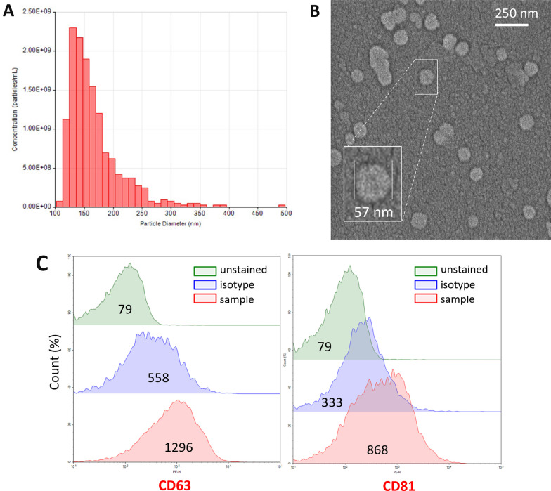
Fig. 5.
Characteristics of the extracellular vesicles released by CSD. EVs isolated from the brain one hour after CSD were evaluated in accordance with MISEV guidelines. A Nanoparticle tracking assay showed that the mean particle diameter was 167 nm, compatible with sEVs (SD = 46.6 nm; d90/d10 = 1.82). The concentration was 1.39e + 10 particles/ml. B SEM image of EVs derived from the mouse brain cortex and a magnified image of a single EV with a 57 nm radius from the same sample (inset). C Flow cytometric analysis of EVs captured with anti-CD63 antibody-coated latex beads and labeled with anti-CD63 and anti-CD81 antibodies verified that they expressed typical EV surface markers. Each sample was compared with an unstained sample and with a sample labeled with an isotype antibody. The numbers in the middle of each histogram show the mean fluorescence intensity

