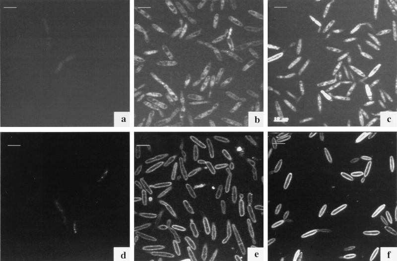
FIG. 6.
Confocal micrographs of U. maydis S023 incubated with 50 μM Fe-B9-LRB (a to c) and Fe-CAT18 (d to f). Fluorescence label can be seen within cells treated with the fluorescent ferrichrome analog (B9-LRB) in comparison with an increase of fluorescence around the cell membranes with the FOB analog CAT18. (a and d) Control; (b and e) after 4 h of incubation; (c and f) after 18 h of incubation. Images were acquired with a Leica TCS4D system and processed by using Adobe Photoshop software on a Power Macintosh computer. Bars, 10 μm.

