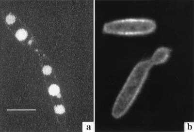
FIG. 7.
Confocal micrographs of U. maydis S023 incubated for 48 h with 50 μM Fe-B9-LRB (a) and Fe-CAT18 (b). Fluorescence derived from the ferrichrome analog (B9-LRB) is clearly concentrated in vesicles within the cell, whereas fluorescence derived from the FOB analog CAT18 is localized around the cell membrane. Images were acquired as for Fig. 6. Bar, 10 μm.

