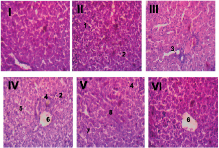
Figure 4.
Histology of the liver. Liver samples of control animals (I) showed well preserved hepatic structural architecture and normal hepatocytes with no observable lesions. Animals in group II (day 1-7 UV maternal exposure group) exhibited liver samples with multifocal hepatocellular degeneration (1) and coagulation necrosis (2). Liver samples from group III animals (day 8-14 UV maternal exposure group) showed moderate centrilobular hepatocellular degeneration. Group IV (day 15-21 UV maternal exposure group) had liver tissues with periportal vacuolar hepatocellular degeneration (5), coagulation necrosis (2), a few foci of inflammation (4) and centrilobular hepatocellular necrosis (6). In group V (day 1-14 UV maternal exposure group), liver samples showed random hepatocellular coagulation necrosis (7), inflammation (4) and fibroblast proliferation (8). Animals in Group VI (day 1-21 UV maternal exposure group) also exhibited centrilobular hepatocellular coagulation necrosis (6).

