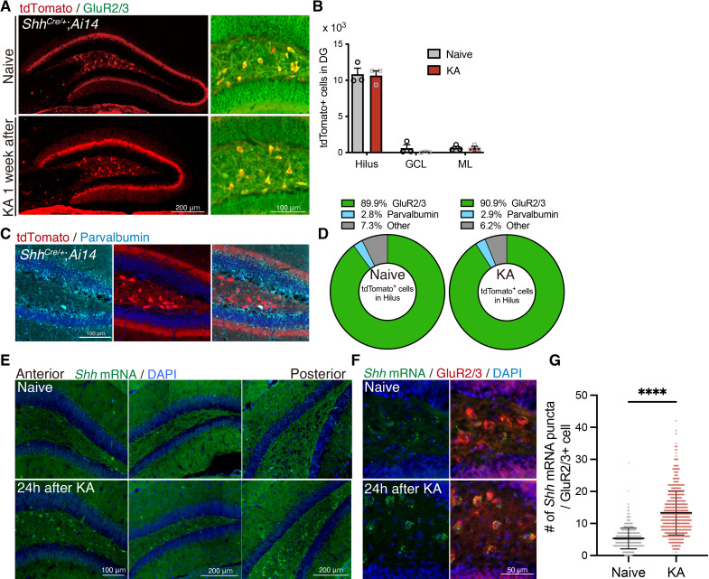
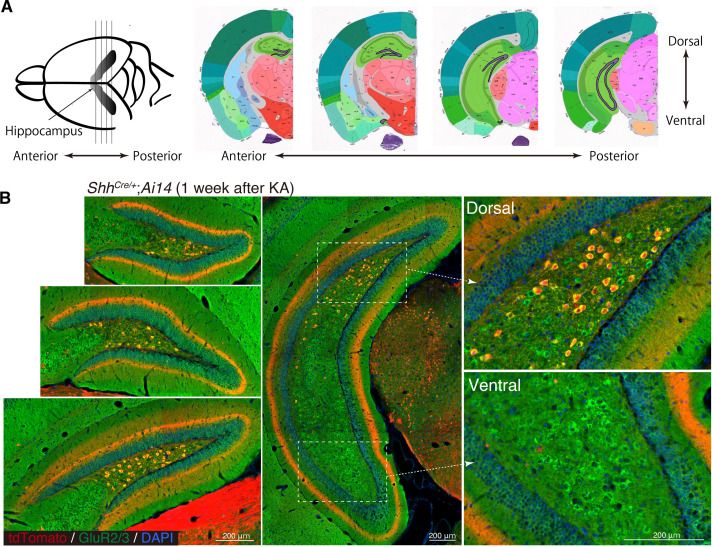
Figure 2. Sonic hedgehog (Shh) is expressed in mossy cells and upregulated upon seizure induction.
(A) Representative immunofluorescence images of mossy cells labeled with GluR2/3 (green) and ShhCre/+;RosaAI14 (tdTomato; red) 1 week after seizure induction. (B) Quantification of tdTomato+ cells in each area of the dentate gyrus (DG). GCL: granule cell layer, ML: molecular layer (naive: n = 3, kainic acid [KA]: n = 3 mice). (C) Representative immunofluorescence images of interneurons labeled with parvalbumin (cyan) and ShhCre/+;RosaAI14 (tdTomato; red) in the hilus. (D) Cell-type population of tdTomato+ cells in the hilus of ShhCre/+;RosaAI14 mice 1 week after seizure induction (naive: n = 3, KA: n = 3 mice). (E) In situ RNAscope detection of Shh mRNA (green) in the DG from anterior to posterior 24 hr after seizure induction. (F) Representative RNAscope-immunofluorescence images for Shh expression in mossy cells labeled with GluR2/3 (red). (G) Quantification of Shh mRNA puncta in GluR2/3+ mossy cells. Values represent mean ± standard error of the mean (SEM); ****p < 0.0001. Unpaired t-test (two-tailed). A total of 608 and 584 GluR2/3+ cells were quantified from three mice in naive and KA treated groups, respectively.


