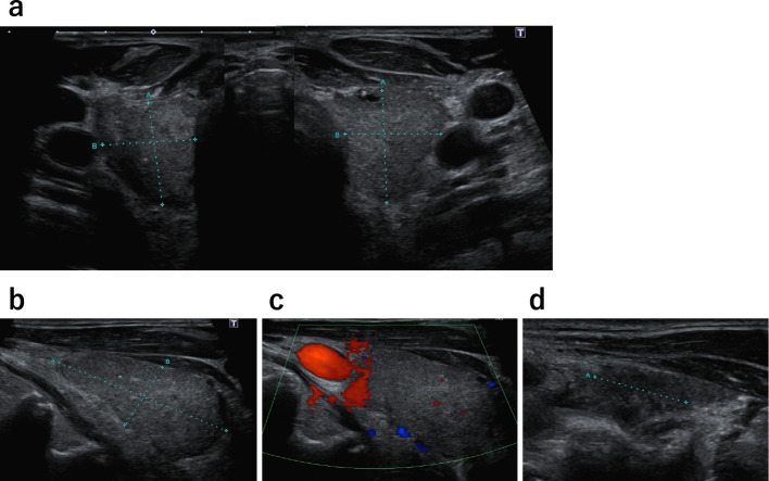
Figure 2.
Thyroid ultrasound. a) The thyroid gland showed no enlargement and presented heterogeneous parenchyma. b) Right thyroid lobe in a sagittal view (41×21×19 mm). c) Thyroid doppler ultrasound showed no hypervascularization. d) Sagittal view of the left thyroid lobe (42×23×17 mm) showed the presence of a 14-mm hypoechoic area. The estimated thyroid volume was approximately 17 g.

