
FIGURE 2.
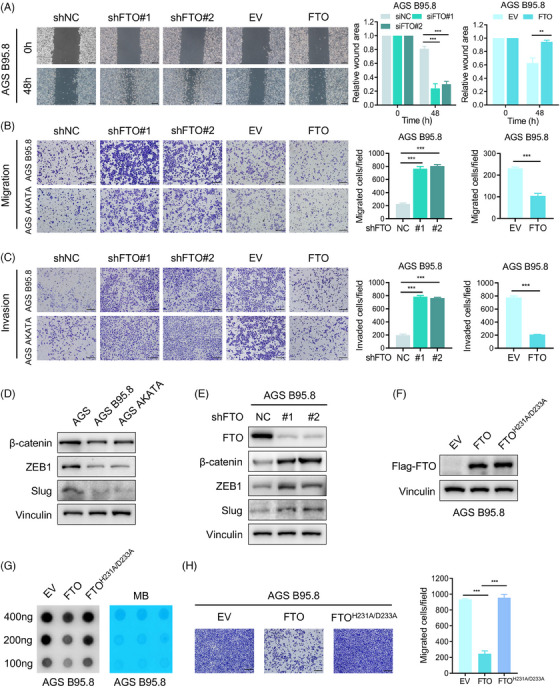
FTO restrains EBVaGC cell migration and invasion in vitro. (A) Wound healing assays of AGS B95.8 cells with FTO silencing and FTO overexpression were recorded and quantitatively analysed. Scale bar: 200 µm. (B and C) Images and quantification of cell migration (top) and invasion (bottom) assays of FTO‐knockdown and FTO‐overexpressing EBVaGC cells. Scale bar: 200 µm. (D) Western blotting analysis was used to detect EMT markers (β‐Catenin, ZEB1, Slug) in EBVaGC and EBVnGC cells. (E) The protein levels of EMT markers (β‐Catenin, ZEB1, Slug) in AGS B95.8 cells after FTO deficiency were measured using immunoblotting. (F) Western blotting assay of FTO in AGS B95.8 cells overexpressing wild‐type and catalytic mutant FTO. (G) RNA m6A dot blot assay of wild‐type and catalytic mutant FTO‐overexpressing AGS B95.8 cells. MB staining served as a loading control. (H) The migration ability of AGS B95.8 cells overexpressing wild‐type and catalytic mutant FTO was determined (left), and the cell migration assay results were quantitatively analysed (right). Scale bar: 200 µm. The data in (A–C and H) are presented as the means ± SDs. p‐Values were determined by Student's t test. *p < 0.05; **p < 0.01; ***p < 0.001. Vinculin was included as a loading control.