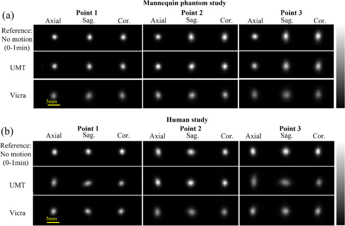
Figure 3.
Reconstructed images of the point sources for (a) the mannequin phantom study and (b) the human volunteer study. Slices shown intersect the highest voxel value for each source for each method. The top row shows the reconstruction of the first minute of acquisition (no motion). The next two rows show the full 15 min reconstructions with motion correction by UIH Motion Tracking (UMT) and Vicra. For each point source, the color bar was scaled to the maximum intensity from the reference image, which differs for each source. All the images for each source were displayed to their respective maxima for visual comparison. UMT: United Imaging Healthcare Motion tracking system.

