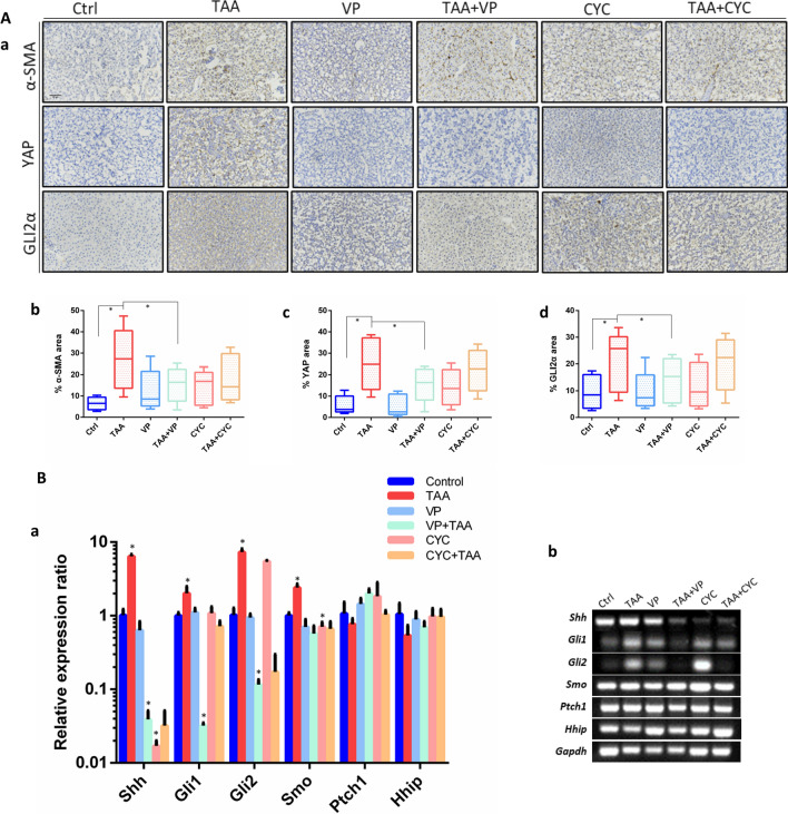
Fig. 5.
Immunohistochemistry detections in zebrafish liver. A (a) Immunostaining of α-SMA, YAP and GLI2α, for the detection of tissue sections of normal control, TAA-induced, VP treatment, TAA + VP treatment, CYC treatment and TAA + CYC treatment. (b–d) Quantitation of α-SMA, YAP and GLI2α immunopositivity by area in the whole liver section in each group. Representative data from 5 slices per group. Scale Bars = 50 μm. B Relative expression of Hh factors in adult zebrafish liver. (a) QRT-PCR test of factors expression. (b) Conventional PCR was as the validation. Data are expressed as the means ± SD. *p < 0.05

