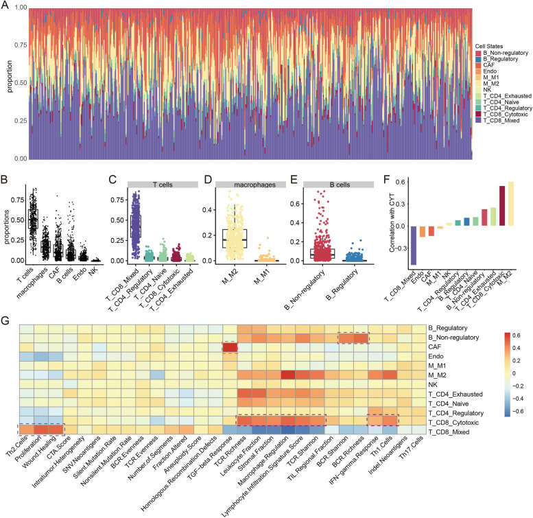
Fig. 3.
Overview of tumor microenvironment (TME) cell states in melanoma samples from TCGA. A Stacked bar charts summarising proportion of each TME cell state in each sample. B-E Dot plots showing the population frequency for each melanoma sample among all TME cell types (B) and among cell states in T cells (C), in macrophages (D) and in B cells (E). F Bar plots showing associations between cell states and cytolytic activity. G Heatmap showing associations between cell states (row) and immune characteristics (column), red for positive correlations and blue for negative correlations. Pearson's correlations were calculated. Dark red boxes highlight the strong positive associations with immune characteristics for the cell states: T_CD8_Cytotoxic, T_CD8_Mixed (Cytotoxic and Exhausted), B_Non-regulatory and CAF

