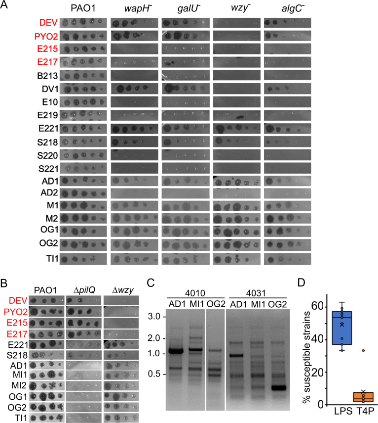
Fig 6.
Phage growth on different P. aeruginosa strains. (A and B) Plating on PAO1 mutants. Phage lysates were diluted (×10) in 96-well plates and replicated on PAO1 and the indicated PAO1 mutants. The plates were incubated overnight at 37°C. wapH −, PAER5b; galU −, PAER6b; wzy −, PADR6; algC −, PAER10b, ΔpilQ, PAMO302; Δwzy, PAMO301. The CK4 components are reported in red. (C) Different RAPD PCR band pattern obtained with primers 4010 and 4031 on the indicated phages. The same patterns were obtained in a replicate experiment. MW marker migration is indicated on the left (kb). (D) Percentage of the P. aeruginosa strains listed in Table S4 susceptible to all phages listed in Table 4 but E221, for which only a subset of clinical strains was analyzed. Each dot represents the host range of a different phage. LPS, phages not growing on wzy mutants; T4P, phages not growing on the ΔpilQ mutant.

