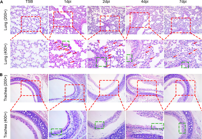
Fig 4.
Histopathology lesions induced in the lungs of mice infected with T. pyogenes. Mice were infected with 2 × 10⁶ CFU of T. pyogenes strain 20121 and sacrificed at 1, 2, 4, and 7 dpi. Samples of (A) lung and (B) trachea were fixed, embedded in paraffin, and sectioned at a thickness of 5 µm. Sections were stained with hematoxylin-eosin and photographed using Zeiss Viewer software; the red box shows greater detail, with extensive inflammatory cell infiltration of the lung (red arrow) and epithelial cell degeneration (green box).

