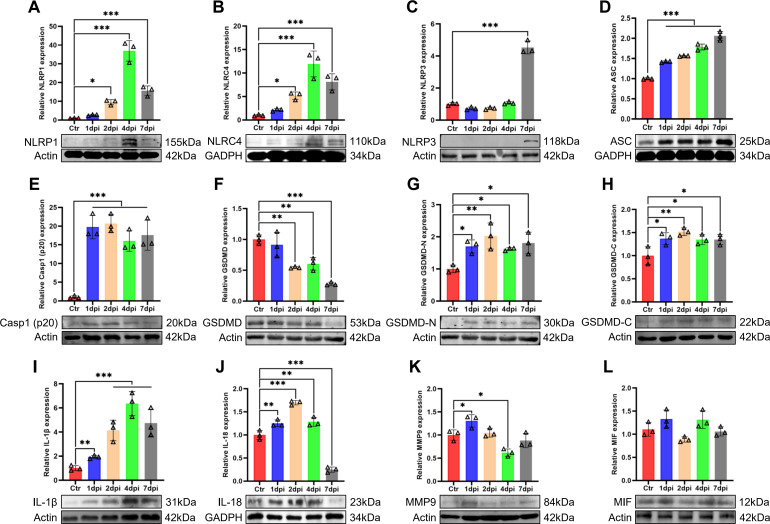
Fig 6.
The NLR/ASC/caspase-1/IL axis is activated in the lungs of T. pyogenes-infected mice. Western blots of T. pyogenes-infected wild-type mouse lungs at 1, 2, 4, and 7 dpi are shown. Relative protein expression of (A) NLRP1, (B) NLRC4, (C) NLRP3, (D) ASC, (E) Casp1(p20), (F) GSDMD, (G) GSDMD-N, (H) GSDMD-C, (I) IL-1β, (J) IL-18, (K) MMP9, and (L) MIF are shown; *P < 0.05, **P < 0.01, and ***P < 0.001. The actin images (F and G) were reused here because they are parts of the same internally controlled experiment from the same gel.

