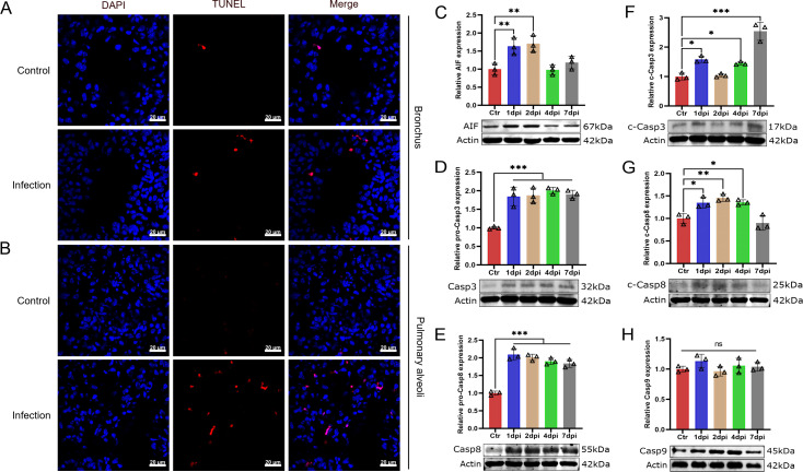
Fig 8.
T. pyogenes infection induces apoptosis in mouse lung cells. (A) Mice were challenged with 2 × 10⁶ CFU of T. pyogenes, and at 2 dpi, lungs were collected for immunofluorescence to verify the occurrence of apoptosis using a TUNEL assay. Apoptosis is shown in red in (A) bronchial epithelial cells and (B) pulmonary alveoli in the lung, and nuclei (DAPI) in blue. Western blot analysis of (C) AIF, (D) caspase-3 (Casp3), (E) caspase-8 (Casp8), (F) cleaved caspase-3 (c-Casp3), (G) cleaved caspase-8 (c-Casp8), and (H) caspase-9 (Casp9) expression shows apoptosis of lung cells at different days of infection. Data are shown as the mean ± SD of three experiments. *P < 0.05, **P < 0.01, and ***P < 0.001. The actin images including panels D and F and panels E and G were reused here because they are parts of the same internally controlled experiments from the same gel, respectively.

