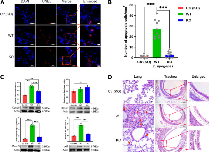
Fig 9.
Comparison of apoptosis and injury induced by T. pyogenes alleviated in the lungs of wild-type and tnf-α-/- mice. WT and tnf-α-/- C57 (KO) mice were challenged with 2 × 10⁶ CFU of T. pyogenes, and at 2 dpi, lungs were collected for immunofluorescence. (A) Apoptotic cells (TUNEL) in trachea epithelial cells were detected with the In Situ Cell Death Detection Kit, and cell nuclei were stained with DAPI. (B) Number of apoptotic cells in trachea epithelial cells from different groups of mice. ***P < 0.001. Protein levels of (C) caspase-8, caspase-9, caspase-3, and AIF were detected by western blot. (D) Comparison of histopathological lesions of lungs and trachea between infected WT and KO mice on 2 dpi, showing the epithelial layer of the respiratory tract of lungs (red dotted boxes) and neutrophils (red arrow).

