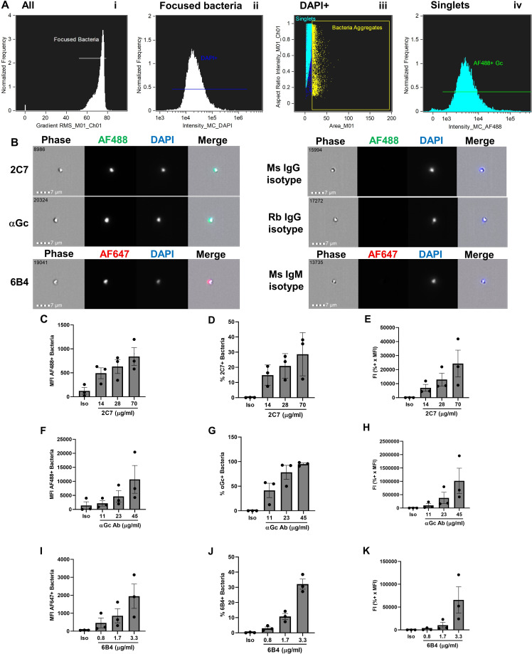
Fig 1.
Antibody binding to the surface of N. gonorrhoeae by imaging flow cytometry. (A) Gating strategy. N. gonorrhoeae strain FA1090 was incubated with rabbit anti-N. gonorrhoeae antibody and AlexaFluor 488 (AF488)-coupled anti-rabbit antibody, fixed, and incubated with DAPI. On the ImagestreamX imaging flow cytometer, samples were gated on (I) Root mean square (RMS) of >52, then (ii) focused bacteria, and then (iii) DAPI+ singlets, from which AF488-positive fluorescence was measured (iv). (B) Representative examples of individual N. gonorrhoeae of strain FA1090 that bind mouse (Ms) 2C7 anti-lipooligosaccharide IgG3, polyclonal rabbit (Rb) anti-N. gonorrhoeae IgG (αGc), and mouse 6B4 anti-lipooligosaccharide IgM (left) but not corresponding isotype controls (right). Number in the upper left corner of each phase image indicates the identity of the particle captured by the imaging flow cytometer. Measurements of mean fluorescence intensity (MFI) (C, F, I), percent-positive bacteria (D, G, J), and fluorescence index (FI = MFI x percent positive) (E, H, K) for N. gonorrhoeae incubated with the indicated concentrations of 2C7 (C–E), αGc (F–H), and 6B4 (I–K) or highest concentration of each isotype control (iso). Bars indicate the mean ± SD of three independent experiments, with each biological replicate as one data point.

