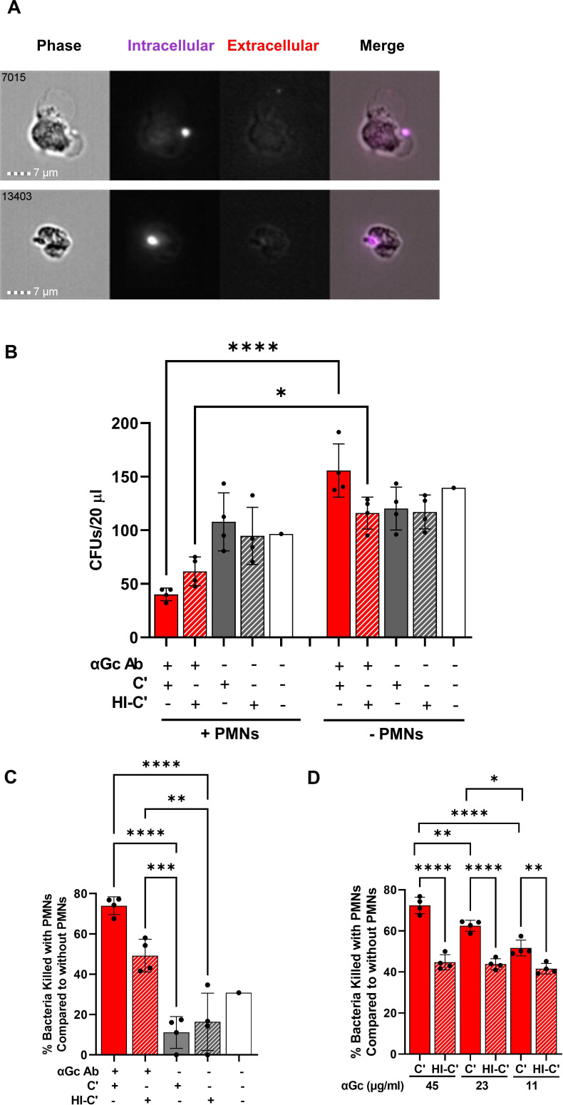
Fig 3.
Opsonophagocytic killing assay with primary human PMNs. (A) Examples of two PMNs that underwent opsonophagocytosis of Tag-IT Violet-labeled (purple) N. gonorrhoeae strain FA1090, in the presence of C6-depleted NHS and rabbit anti-N. gonorrhoeae antibody (50 µg/mL), analyzed by imaging flow cytometry. PMNs were fixed and stained with Alexa Fluor 647-coupled anti-N. gonorrhoeae antibody to detect bound but not phagocytosed bacteria (red). The number in the upper left corner of each phase image indicates the identity of the cell captured by the imaging flow cytometer. (B) N. gonorrhoeae strain FA1090 was mixed with rabbit anti-N. gonorrhoeae antibody or buffer control as the above, except not fluorescently labeled. C6-depleted normal human serum that was untreated (C’) or heat inactivated (HI-C’) was added to 5% final concentration, along with primary human PMNs (+PMNs) or buffer (−PMNs). CFUs were enumerated from 20 µL of the incubation mix. Red solid bar with PMNs is the full opsonophagocytic condition. (C) Results are as in B except each condition was expressed as a percentage of the enumerated CFU for the same condition in the absence of PMNs. (D) Results are as in B except with the indicated concentrations of rabbit anti-N. gonorrhoeae antibody and presented as the percent of bacteria killed compared with bacterial killing when heat-inactivated serum was used. In B–D, results presented are the average ± SD from four independent experiments using different human subjects’ PMNs, with each biological replicate as one data point. *P ≤ 0.05, **P ≤ 0.005, ***P ≤ 0.001, and ****P ≤ 0.0001 by one-way ANOVA followed by Holm-Šidák multiple comparisons test.

