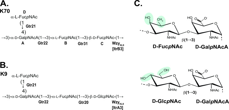
Fig 3.
Comparison of CPS and sugar structures. (A) Structure of K70 CPS from A. baumannii SGH0807 (this work). (B) Structure of K9 CPS from A. baumannii LUH3484 (39). Glycosyltransferases are shown in bold next to the linkage they have been assigned to. (C) Representation of the glycosidic linkages formed by WzyKL9 shown to the right of each CPS structure with differences highlighted by colored shading.

