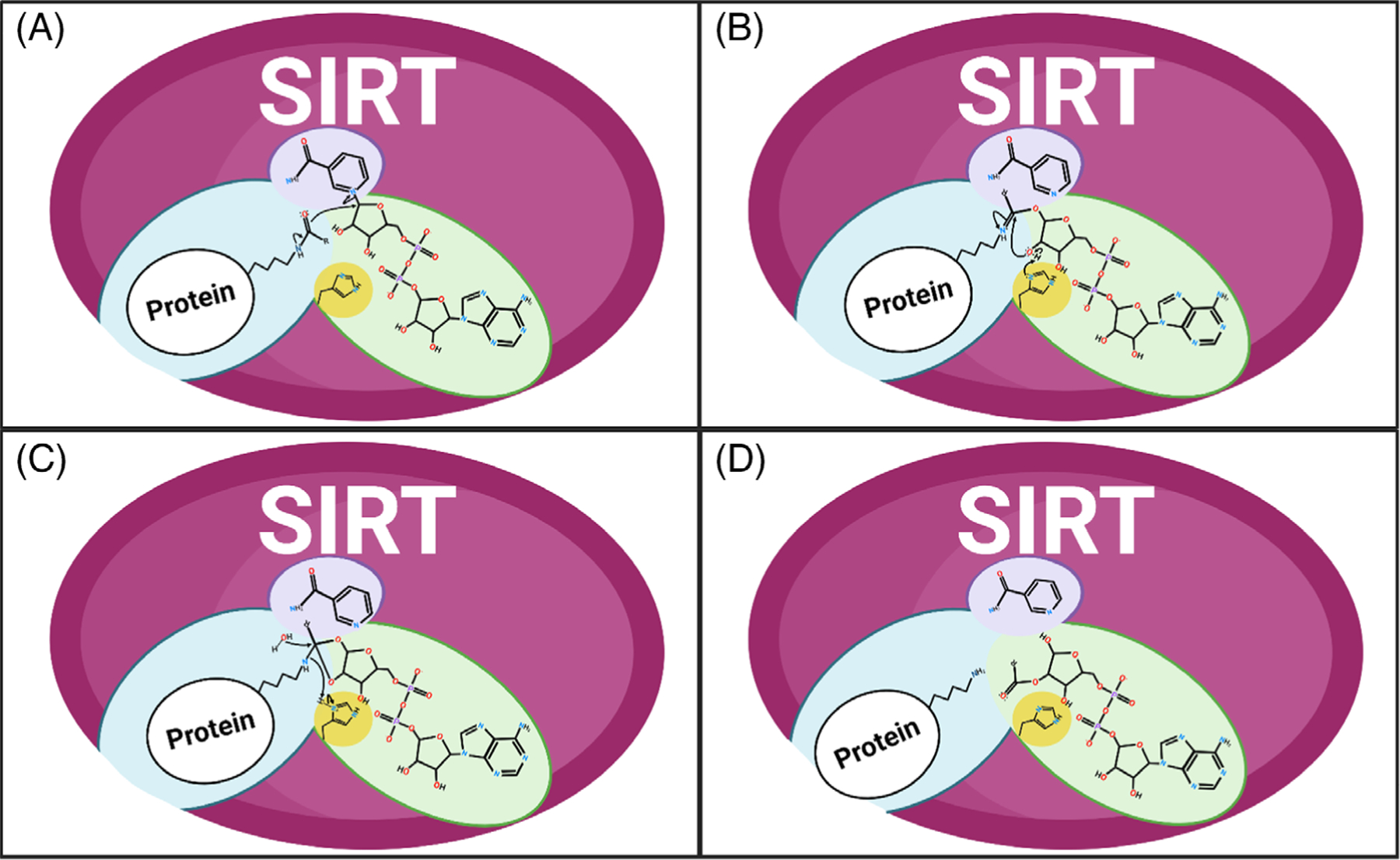
FIGURE 4.

Sirtuin enzymatic deacylation of substrate. (A) Nicotinamide cleavage. Nicotinamide adenine dinucleotide (NAD+) is shown in the purple and green pockets of the representative SIRT enzyme. Acylated lysine protein substrate is highlighted in the blue pocket. (B,C) Formation of the alpha-1’-O-alkylamidate intermediate. Nicotinamide is shown in the purple pocket, the alpha-1’-O-alkylamidate intermediate in the blue and green pockets, and the SIRT catalytic histidine in the yellow pocket. (D) Deacylation of lysine. The reaction yields deacylated lysine (blue pocket), nicotinamide (purple pocket), and 2’-O-acetyl-adenosine diphosphate ribose (green pocket).
