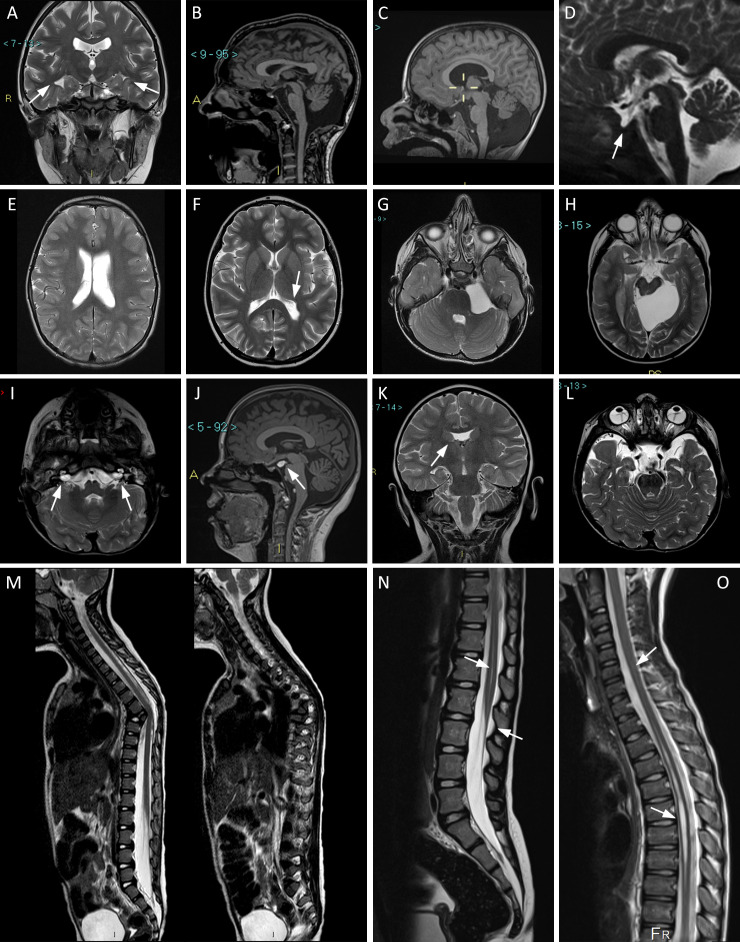
Figure 1.
Representative MRI images in our cohort. Arrows indicate the relevant details. (A) Pt. 16, coronal T2-weighted view: incomplete hippocampal inversion, bilateral. (B) Pt. 7, sagittal T1: large cisterna magna. (C) Pt. 4, sagittal T1: Dandy-Walker variant. (D) Pt. 25, sagittal T2: empty sella. (E) Pt. 23, axial T2 TSE: dilated and asymmetric lateral ventricles. (F) Pt. 10, axial T2 TSE: left trigonal enlargement. (G) Pt. 5, axial T2: arachnoid cyst in left cerebellopontine angle. (H) Pt. 31, axial T2: arachnoid cyst in left ambient cistern. (I) Pt. 12, axial T2: bilateral patulous internal auditory canal. (J) Pt. 16, sagittal T1: lipoma tuber cinereum. (K) Pt. 3, coronal T2: partial agenesis of septum pellucidum. (L) Pt. 12, axial T2: bilateral persistent hyperplastic primary vitreous. (M) Pt. 52, sagittal T2: focal dorsal kyphosis due to anomalous thoracic T6-T7 vertebral differentiation. (N) Pt. 51, sagittal T2 TSE: hydromyelia and low-lying Conus medullaris. (O) Pt. 38, sagittal T2 TSE: hydrosyringomyelia. Pt., participant.

