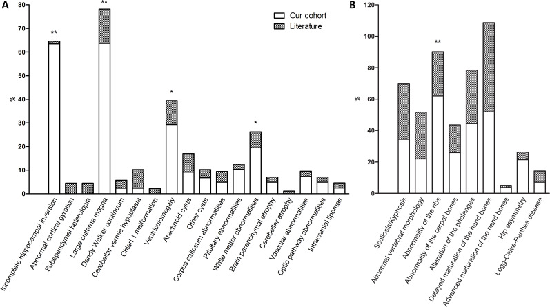
Figure 4.
Stacked histograms comparing the imaging findings in our cohort with those reported in the literature. (A) Brain abnormalities at diagnostic imaging. Cortical gyration anomalies consist solely of the four mild alterations reported in a volumetric study, and no macroscopic gyration defects were detected by standard MRI in any of the other publications. (B) Skeletal abnormalities at diagnostic imaging. Skeletal features mainly determined through physical examination rather than X-ray imaging, such as wide anterior fontanel and hip dysplasia, were not included because of the difficulty of establishing accurate ratios of evaluated patients. *Significant difference at p<0.05. **Significant difference at p<0.001.

