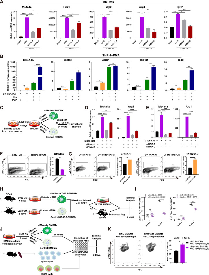
Figure 2.
MS4A4A promotes M2 macrophage polarisation and induces CD8+ T-cell dysfunction. (A) BMDMs were transfected with Ms4a4a-specific siRNA or negative control siRNA and polarised into the M2-phenotype using IL-4 (20 ng/mL) and IL-13 (20 ng/mL). The interference efficiency of Ms4a4a and the expression of M2 markers (Fizz1, Mgl2, Arg1 and Tgfb1) were measured by qRT-PCR. (B) An MS4A4A-overexpressing cell line was constructed using the human monocyte cell line THP-1, and then the engineered THP-1 cells were induced to differentiate into M0 macrophages with PMA (50 ng/mL). IL-4 (20 ng/mL) was used to stimulate M0 macrophages to polarise into the M2-phenotype, and the expression levels of MS4A4A and M2 markers (CD163, VEGFA, IL-10, ARG1 and TGFB1) were measured by qRT-PCR. (C–G) Study of the effect of MS4A4A expression on macrophage polarisation in vitro. (C) Bone marrow cells from C57BL/6 mice were extracted in vitro and induced into BMDMs using L929 cells conditioned medium (L929-CM). The BMDMs were then transfected with MS4A4A-specific siRNA or control siRNA on day 6. After 48 hours, the macrophages were cultured in MC38 cells conditioned medium (MC38-CM) or CT26 cells conditioned medium (CT26-CM) for 24 hours. (D–E) The expression levels of Ms4a4a and Arg1 were measured by qRT-PCR (n=3). (F–G) The proportion of M2 macrophages in each group of macrophages was detected by flow cytometry (n=3). (H–I) In vivo confirmation of the effect of MS4A4A on the M2 polarisation function of TAMs. (H) The BMDMs with MS4A4A differential expression (siNC-CD45.2 and siMs4a4a-CD45.1) were labelled with CFSE, and then the two types of cells were mixed at a ratio of 1:1. Some of these cells were cultured in vitro, and others were transferred into tumour-bearing mice. After 3 days, FACS was performed on the above donor cells. (I) FACS analysis of CD206 and IL-10 expression in two types of donor cells (siNC-CD45.2 and siMs4a4a-CD45.1) in vitro and in vivo (n=5). (J–K) Study of the effect of macrophage MS4A4A expression on CD8+ T-cell function in vitro. (J) Experimental design. (K) FACS analysis of Ki67 expression on the indicated CD8+ T cells (n=3). Results are represented as mean±SEM. BMDMs, bone marrow-derived macrophages; CFSE, carboxyfluorescein succinimidyl ester; CM, conditioned medium; FACS, flow cytometry; FSC, forward scatter; IL, interleukin; LV, lentiviral vectors; mRNA, messenger RNA; PMA, phorbol 12-myristate 13-acetate; qRT-PCR, quantitative real-time PCR; siMs4a4a, MS4A4A-specific siRNA; siNC, control siRNA; siRNA, small interfering RNA; TAMs, tumour-associated macrophages.

