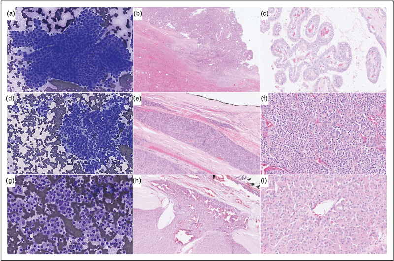
FIGURE 1.
Cytological and histopathological features of follicular-derived thyroid neoplasms. (a–c) Invasive encapsulated papillary carcinoma. Cytological smear shows typical elongated nuclei with grooves and pseudoinclusions leading to Bethesda VI classification (a) (Diff-Quick staining, X200). Transcapsular invasion (b) and papillary architecture with nuclear score of 3. (c) [HE staining, X200 (b), X400 (c)]. (d–f) Angioinvasive poorly differentiated thyroid carcinoma. Cytological smear shows highly cellular crowded groups of uniform follicular cells arranged in microfollicles Bethesda IV (d) (Diff-Quik staining, X200). Vascular invasion (e) and solid/trabecular and microfollicular architecture (f) [HE staining, X200 (e), X400 (f)]. (g–i) Oncocytic follicular thyroid carcinoma. Cytological smear shows highly cellular groups of oncocytic cells Bethesda IV (g) (Diff-Quik staining, X200). Transcapsular invasion (h) and oncocytic cells arranged in a microfollicular architecture (i) [HE staining, X200 (h), X400 (i)].

