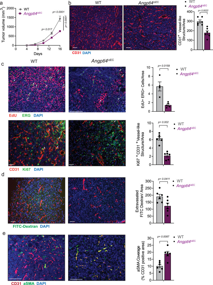
Fig. 2. EC-specific deletion of Angptl4 reduces adult pathological angiogenesis in vivo.
a–e Tumor analysis of WT (n = 6) and Angptl4iΔEC (n = 9) mice with s.c. injection of LLCs in the dorsal flank. a LLC isograft progression (tumor volume) in WT and Angptl4iΔEC mice. b Left, representative micrographs of CD31 (red) and DAPI (blue) immunostaining. Right, quantification of CD31+ vessel-like structures. c Upper left, representative micrographs of immunofluorescence staining for EdU (red), and ERG (green), DAPI (blue). Proliferating (EdU and ERG double-positive) ECs are shown in yellow (arrow). Upper right, quantification of EdU and ERG double-positive cells (n = 4 for each genotype). Lower left, representative micrographs Ki67 (green) and CD31 (red) and DAPI immunostaining. Lower right, quantification of Ki67 and CD31 double positive cells shown in yellow (arrow) (n = 6 for each genotype), d Left, representative images of FITC-Dextran (70 kDa) (green), CD31 (red) and DAPI immunostaining. Right, quantification of extravasated FITC-Dextran. e Left, representative micrographs of αSMA (green), CD31 (red) immunostaining. Right, quantification of αSMA covered CD31+ vessel-like structures. Right panels (b–e) quantification of at least 4 different images from each mouse, (n = 6 quantified per genotype out of 6 or 9 randomly selected, except for c (n = 4 or 5 per genotype were used). Scale bars, 70μm (b, d, and c lower panel), 125μm (e), 200μm (c upper panel). Data are represented as means ± SEM. Two-way ANOVA with Tukey’s multiple comparisons test in (a) and Mann–Whitney U test in (b–e). Exact p values are shown for each comparison. Source data are provided as a Source data file.

