
Figure 5. Reductions in type 2 serum and tissue biomarkers as a function of dupilumab treatment.
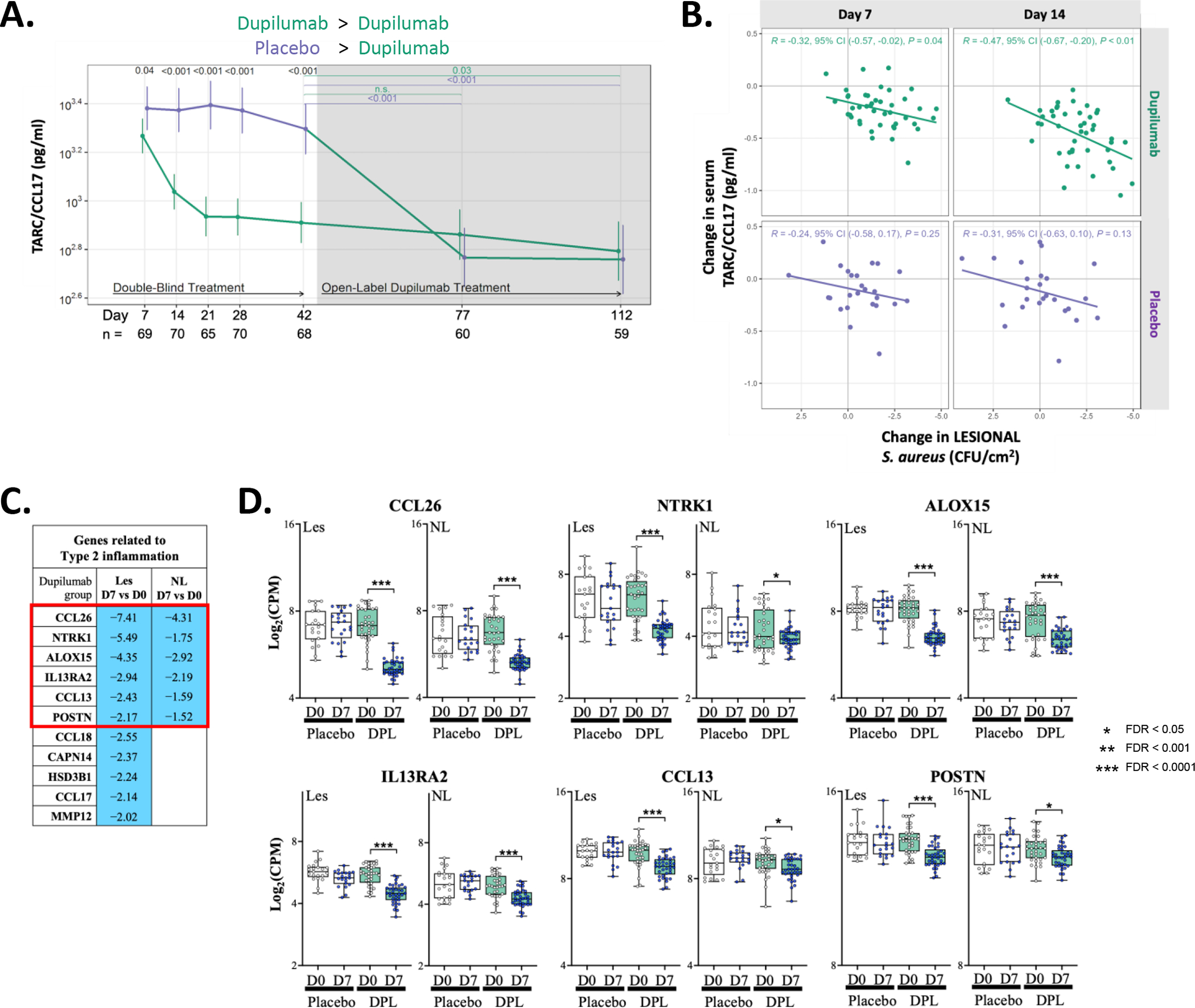
(A) Longitudinal changes in serum TARC/CCL17 during RDBPC and OLE phases of the study. The number of participants with evaluable data at each timepoint are noted below the X axis (with the total population denoted in black, dupilumab-randomized participants in green and placebo-randomized in purple). (B) Reductions in serum CCL17 significantly correlated with reductions in S. aureus lesional CFU abundance at both day 7 (p=0.04) and day 14 (p=0.01) in dupilumab-treated participants but there was no correlation observed at either timepoint in placebo-treated participants. (C) Differentially expressed genes (DEGs) related to type 2 inflammation (with a FDR of <0.05 and with a fold change > 2.0 in lesional skin) is shown comparing day 7 to day 0 in the dupilumab-treated group. Blue indicates reduced expression at day 7 in the dupilumab-treated group. (D) Graphical illustration of the genes reduced in both Les and NL skin (Red Box in [C]). Placebo data is shown in open box plots and dupilumab in shades of green.
CFU, colony forming units; FDR, false discovery rate; les, lesional skin; NL, nonlesional skin