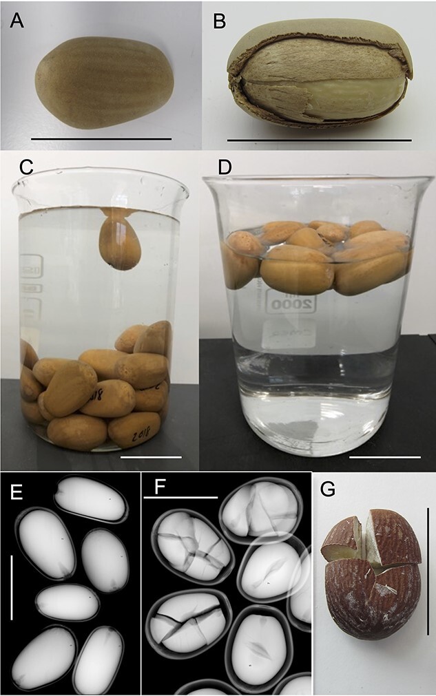
Figure 1.

A) Intact sclerotesta of M. fraseri following manual removal of the outer pulpy sarcotesta using a rotating cement mixer with crushed rock aggregate and water; B) Longitudinally dissected sclerotesta showing the location and structure of the underlying thin membranous endotesta and the inner white megagametophyte; C) 30-month-old seeds stored under standard seed bank conditions (~20% RH and 5°C) subjected to a floatation test for 5 min; D) 54-month-old seeds stored under standard seed bank conditions subjected to a floatation test for 5 min; E) X-ray image of seeds stored for 6 months showing fully formed megagametophyte tissue within each seed with minimal signs of shrinkage or damage; F) X-ray image of seeds stored for 66 months showing fragmentation and significant megagametophyte shrinkage and G) Shattered megagametophyte following extraction from the sclerotesta as observed in Figure 1F. Line = 40 mm.
