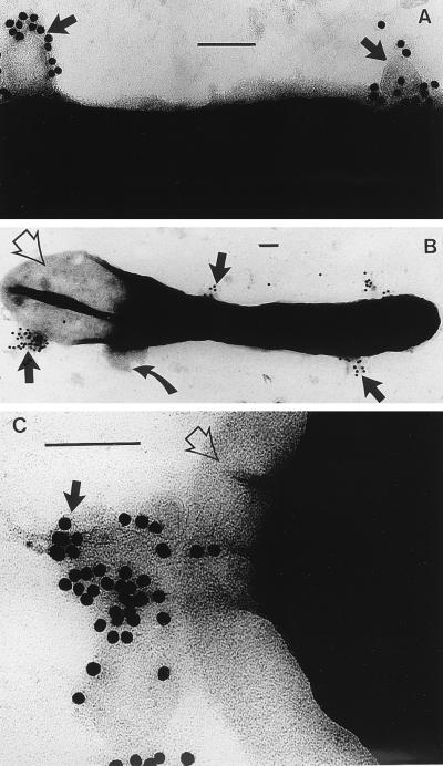
FIG. 2.
(A) Negative stain of the surface of B. stearothermophilus PV72/p6 immediately after MVs have been added to the cells. The MVs have been labelled with an anti-B-band LPS immunogold which is specific for the MVs (14); the MVs have attached to the surface of the bacterium (arrows). The magnification bar in this and the following panels represents 100 nm. (B) An entire B. stearothermophilus cell after 10 min of MV treatment is shown. Large membranous vesicles (those not labelled with colloidal gold), derived from the bacterium’s plasma membrane, are seen extruding from the cell where the peptidoglycan has been hydrolyzed (curved arrow). One pole of the bacterium has been emptied of cytoplasm (empty arrow). MVs (labelled with gold) are still attached to the bacterium (solid arrows). (C) High magnification of the pole of a cell which is extruding cytoplasm since the peptidoglycan layer has been hydrolyzed (empty arrow). MVs and the gold label still remain attached (black arrow).

