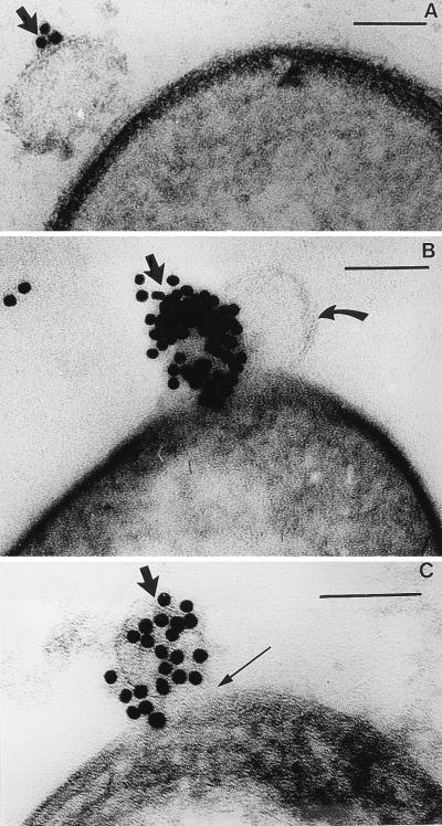
FIG. 3.
(A) Thin section of B. stearothermophilus PV76/p2 showing the initial stage of MV attachment to the bacterial surface. The arrow points to a gold-labelled MV. The magnification bar in this and the following panels represents 100 nm. (B) The MV in this image has lysed the cell after 20 min, and a balloon of cytoplasm, bounded by the plasma membrane (curved arrow), is extruding from the cell. The plasma membrane is not labelled, but an adjacent MV (solid arrow) is labelled by its immunogold probe. (C) In this image, the gold-labelled MV (thick arrow) has completely hydrolyzed the peptidoglycan layer (thin arrow) after 30 min.

