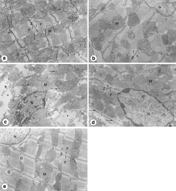
Fig. 3.

The ultrastructure of cardiomyocytes from the left ventricular anterior free wall at the level of the near apex. a Group C showed finely dispersed nuclear chromatin, slightly contracted myofibrils (F), well arranged Z disk (Z), mitochondria (M) with tightly packed cristae, and an intact sarcolemma. b Group EP showed results similar to those described in group C. c Group ISO showed aggregated chromatin of the nucleus (N), disarrayed myofibrils, swollen mitochondria with obvious amorphous matrix densities (arrows) and disorganized cristae, ruptured sarcolemma, and edema of sarcoplasm (asterisk). d Group EP + ISO showed some aggregated chromatin, stretched myofibrils with prominent I-bands (I), slightly swollen mitochondria with cristae visible but not packed, and well preserved sarcolemma with only occasional subsarcolemmal blebs (arrows). e Group CHE + EP + ISO showed finely dispersed nuclear chromatin, well organized myofibrils, slightly swollen mitochodria with disarrayed cristae, and increased dilation of sarcoplasmic reticulum (D). Original magnification, ×10,000
