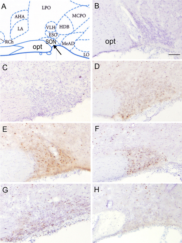
Fig. 1.

Representative sections showing c-Fos expression in the supraoptic nucleus (SON) induced by different RWIS durations. a The exact location of the SON. b The specificity of the immunostaining verified by incubation of the brain sections of group 60 rats with normal goat serum. c There were very few c-Fos-positive neurons in the SON in group 0, but there were many prominent c-Fos-positive neurons in the SON in groups 30 (d), 60 (e), 120 (f), and 180 (g), and the expression of c-Fos tended to be maintained at a higher level during the stress. h There were few c-Fos-positive neurons in the SON in group 30-30. In the whole panel, brownish oval or round spots are the cellular nuclei of the c-Fos-positive neurons, and blue spots are the cellular nuclei of the c-Fos-negative neurons. Scale bars 100 μm. opt Optic tract
