
Fig. 1.
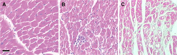
Isoproterenol-induced histological changes. Sections of paraffin-embedded hearts from a untreated, control mice and from mice b 24 h and c 4 weeks after isoproterenol injection were stained with haematoxylin and eosin for nuclei and cytoplasm, respectively. Myocardial injury is suggested by increased cellular infiltration reflected in nuclear aggregation, as in (b), and the presence of disrupted myocardial structure, as in (c). Bar 10 μm