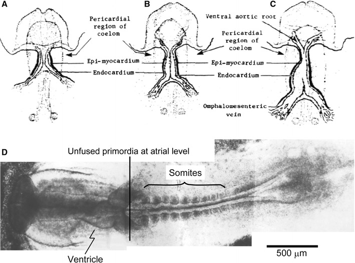
Fig. 1.

a–c Diagrammatic illustration of ventral view to show the origin and subsequent fusion of the paired primordia of the chick heart. a The 6- to early 7-somite stage; b the early 8-somite to early 9-somite stage; and c the 9-somite to 10-somite stage. d Photomontage of the ventral view of a chick embryo with 9 pairs of somites. Modified from Ref. [32]
