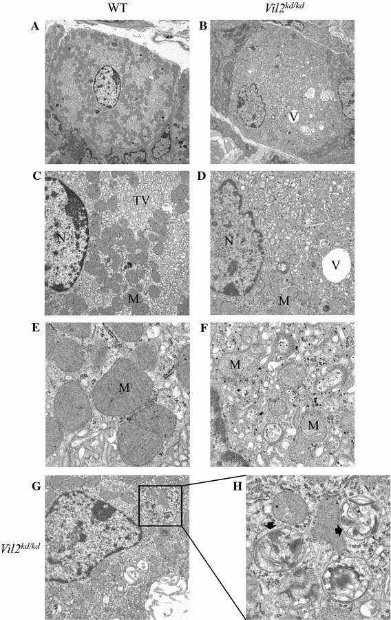
Fig. 7.

Ultrastructure of parietal cells of wild-type (a, c, e) and Vil2 kd/kd (b, d, f) mice at different magnifications. a, b Low magnifications (×3610) of parietal cells. c–f Higher magnifications of parietal cells (c, d ×9300; e, f ×31,800). g, h Abnormal multilamellar structures (arrows) were observed in the parietal cells of Vil2 kd/kd mice at low (×9300) (g) and high (×31,800) (h) magnification. N nucleus, M mitochondrion, TV tubulovesicle, V vacuolar structure
