
Fig. 1.
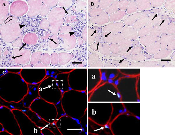
a Hematoxylin and eosin staining 3 days after ECC. Damaged myofibers were defined as those inflammatory cells with swollen (outlined arrows), swollen with infiltration (arrows), and infiltration (arrow-heads) appearance. b The multiple central nuclei can be detected (arrows) after 7 days of ECC. c TUNEL-positive nuclei were found both inside (a) and outside (b) myofibers. Muscle transverse sections were immunofluorescent-stained with dystrophin antibody to identify the sarcolemma (red), fluorescein-mediated TUNEL assay was performed to identify apoptotic nuclei (light blue), and all nuclei were labeled by DAPI staining (blue). TUNEL-positive nuclei positioned inside the dystrophin stain (a) were identified as myofiber nuclei, whereas other nuclei located outside (b) were counted as endothelial or interstitial cell nuclei. Bar 50 μm