
Fig. 2.
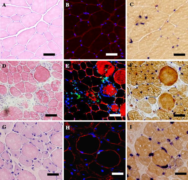
Serial transverse sections from control (a–c), 3 days (d–f), and 7 days (g–i) after ECC in TA muscle. H&E staining (a, d, g) was performed to examine the histological features of muscle damage, triple histochemical staining (b, e, h) for myofiber apoptotic nuclei was used as shown in Fig. 1, and AP-DPPIV staining was performed to identify endothelial cells (c, f, i). Bar 50 μm