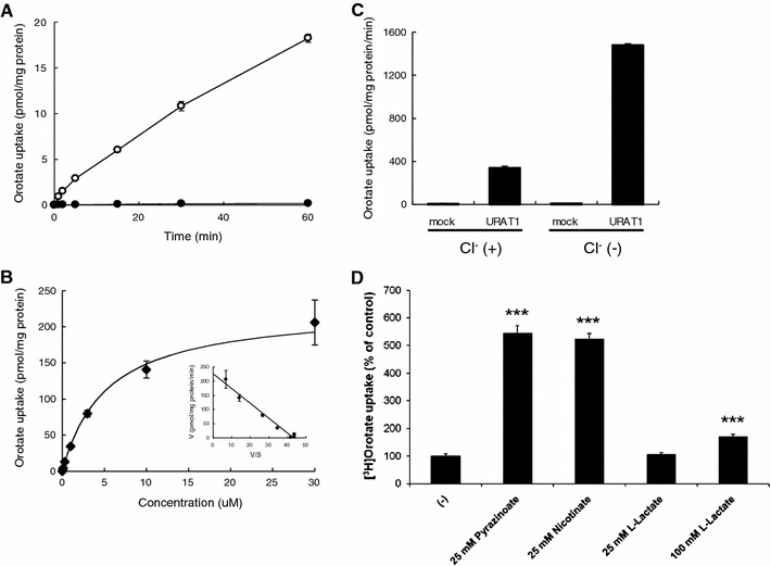
Fig. 1.

Orotate uptake by hURAT1. a Time course of orotate uptake in HEK-hURAT1 cells. hURAT1 and mock cells were incubated in Cl−-free HBSS solution containing 50 nM [3H]orotate for 2 min at 37°C. Open circles represent uptake into hURAT1-expressing cells. Closed circles represent uptake into mock cells (mean ± SEM; n = 4). b Concentration-dependent uptake of orotate in HEK-hURAT1 cells. Uptake of 50 nM [3H]orotate by control or hURAT1-expressing cells was measured for 2 min at various concentrations (0.03, 0.1, 0.3, 1, 3, 10, 30 μM) (mean ± SEM; n = 4). Inset, Eadie-Hofstee plot. V, velocity; V/S, velocity per concentration of orotate. c Orotate uptake via hURAT1 was stimulated by removal of Cl− from the bath. Transport rates of [3H]orotate (50 nM) in HEK-mock and HEK-hURAT1 cells were measured for 2 min in the presence or absence of extracellular Cl−. Each value represents the mean ± SE of four monolayers from two separate experiments. d Trans-stimulatory effect of test anions on the uptake of [3H]orotate via hURAT1. Control and hURAT1-expressing oocytes were injected with several unlabeled anions indicated, or water and incubated for 15 min at RT. Then, the oocytes were incubated with [3H]orotate (50 nM) and its uptake for 1 h was determined. The data are mean ± SEM with n = 6–8. ***P < 0.001 versus the uptake of water injected oocytes
