
Fig. 3.
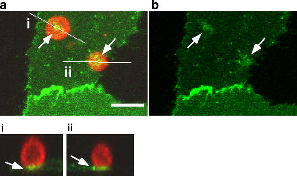
Involvement of PECAM-1 in monocyte adhesion at peri-junctional sites. Adhesion of THP-1 cells to ECs at peri-junctional sites induced a clear cluster of endothelial PECAM-1 at the adhesion site (arrows). Red THP-1 monocytes. Green PECAM-1-GFP in ECs. a The merged image of red and green channels. b The green channel. Maximum-projected xy-images toward the z-axis are shown with orthogonal images (i and ii) cut with the specified planes that are indicated as white lines on xy-images. Bar 20 μm