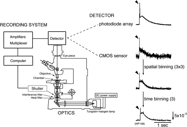
Fig. 1.

Schematic drawing of the optical recording system and optical signals induced by olfactory nerve (N.I) stimulation in a 12-day embryonic chick olfactory bulb. These signals were detected at the same region in one preparation using a 464-element photodiode array (NeuroPDA, RedShirtImaging, Fairfield, CT, USA), or a 128 × 128-element CMOS sensor (NeuroCMOS, RedShirtImaging, Fairfield, CT, USA) with a single sweep. One element of the photodiode array collects light from a round area (diameter = 46 µm), whereas that of the CMOS sensor collects light from a 12.5-µm2 square region. The frame rate of the photodiode array is almost the same as that of the CMOS sensor (≈1 kHz). To increase the S/N, the optical signal detected using the CMOS sensor was processed with spatial binning (average of 3 × 3 elements) and time binning (average of three points). The direction of the arrow in the lower right indicates an increase in transmitted light intensity (a decrease in dye absorption), and the length of the arrow represents the stated value of the fractional change. Arrowheads indicate spike-like signals corresponding to action potentials
