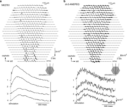
Fig. 2.

Multiple-site optical recording of neural responses to upper spinal cord stimulation in 7-day embryonic chick brainstem-spinal cord preparations. The preparation was stained with a merocyanine-rhodanine absorption dye, NK2761 (a), or a styryl fluorescence dye, di-2-ANEPEQ (b). The optical signals evoked by electrical stimulation (200 µA/1 ms) were recorded simultaneously from 464 contiguous regions of the preparation with a magnification of ×4 (an objective) ×1.67 (an eyepiece). Electrical stimulation elicited a propagating depolarization wave, widely propagating correlated activity, in the spinal cord and brainstem. Illustrations of the preparations are shown in the lower right, in which the detected areas are marked with gray hexagons. The recording was made in a single sweep. For each recording, enlarged traces of the optical signals detected from four regions indicated by 1–4 are shown in the lower panels. Modified from Ref. [24]
