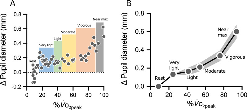
Fig. 2.
A Typical example of the change in pupil diameter. B Change in pupil diameter compared to rest. The data for each intensity are plotted for %. (x-axis) and Δ pupil diameter (y-axis). The plots from left to right show rest, very-light, light, moderate, vigorous, and near maximal/maximal. Uncorrected pupil diameter data were used for statistical analysis. Data are presented as mean. The gray bands represent 95% CI, although some are too small to clearly visualize. The horizontal error bars (95% CI) cannot be visualized as all were smaller than 1.0%

