
Fig. 1.
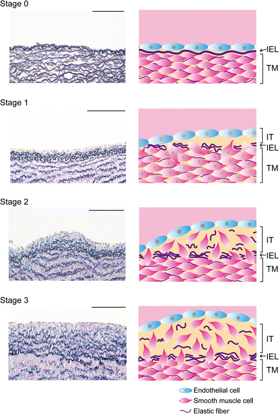
Staging of IT in sheep DA. The left column demonstrates the elastica van Gieson staining of singleton sheep fetuses. All parts of the DA of sheep at 82 days gestation exhibited a single internal elastic lamina and no IT formation (stage 0). The DA of sheep at 100 days gestation contained a part showing the duplication or partial disruption of internal elastic laminae with very modest IT formation (stage 1). The DA of sheep at 103 days gestation showed modest IT formation accompanied by apparent SMC migration (stage 2). All parts of the DA at 147 days gestation exhibited prominent IT formation (score 3). The right panels show schematic models corresponding to histological changes in each stage. IEL internal elastic lamina, TM tunica media, IT intimal thickening. Scale bars 100 µm