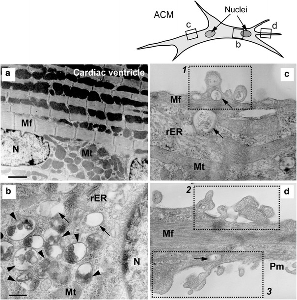
Fig. 4.

Ultrastructures of cardiac ventricle and ACMs. TEM images of mouse cardiac ventricle (a, ×6,000; bar 1.7 µm) and various parts of ACM cultured for 5 days (b–d; ×25,000; bar 400 nm). b–d the enlargement of the small areas of corresponding squares on a line drawing of the cell (upper right). N nucleus, Mt mitochondria, Mf myofiber, rER rough endoplasmic reticulum, Pm plasma membrane. Arrowheads and arrows indicate autophagic bodies containing degraded multiple organelles and almost empty vacuoles, respectively. Dotted square (1–3) the plasma membrane ruffles
