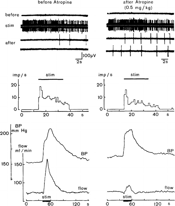
Fig. 12.

Response of vasodilator fibers to the skeletal muscle to electrostimulation within the defense area as an essential component of the defense reaction. Further shown are accompanying changes in arterial pressure (BP) and blood flow measured in the lumbar aorta (flow, electromagnetic flow probe). Recording traces and diagrams without atropine treatment (left) and atropine treatment to block muscarinic cholinergic transmission (right). Upper 4 traces show activity in a sympathetic filament innervating skeletal muscle in which no spontaneous activity exists during the pre-stimulation phase (before). During stimulation (stim) the filament shows strong activation with some decrement in the course of stimulation, and after stimulation (after, two subsequent traces covering ≈40 s) activity returns to silence more or less rapidly. For the diagrams below, the period of electrostimulation within the defense area is indicated by horizontal bars (stim). Middle diagrams represent the discharge rates of the recorded vasodilator fibers being silent before stimulation and becoming silent again with some delay after the end of stimulation. Lower diagrams show stimulus-induced comparable rises of arterial pressure without (left) and with (right) cholinergic blockade with atropine. Stimulus-induced blood flow increase in the lumbar aorta (left) was almost completely suppressed after atropine treatment (right), whereas vasodilator fiber activity was not affected (upper right-hand recording traces). From Horeyseck et al. [21], modified
