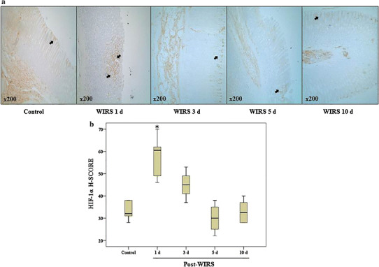
Fig. 2.

a Immunohistochemical labeling of HIF-1α in gastric tissues during gastric lesion healing in rats exposed to WIRS for 6 h. Original magnification ×200. b Graphics of the mathematical values of the H-SCORE evaluations of HIF-1α in gastric tissues. Data are presented as box plots showing the range, quartiles (25–75 %) and median. *p < 0.05 vs. the control group (n = 6 rats/group)
