
Fig. 1.
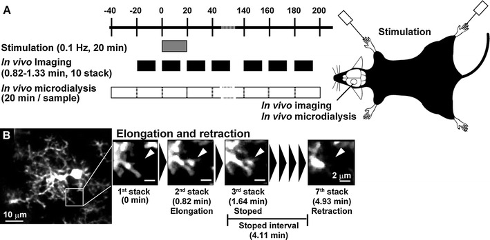
a Schematic drawing of the experimental protocol. In vivo imaging and in vivo microdialysis sampling were carried out every 20 min before and after vibrotactile limb stimulation (0.1 Hz, 20 min). Imaging and microdialysis sampling were carried out on the left somatosensory cortex. b In vivo images of a single microglia (left panel) and representative images of a process showing elongation, retraction, and temporary stop (right panels)