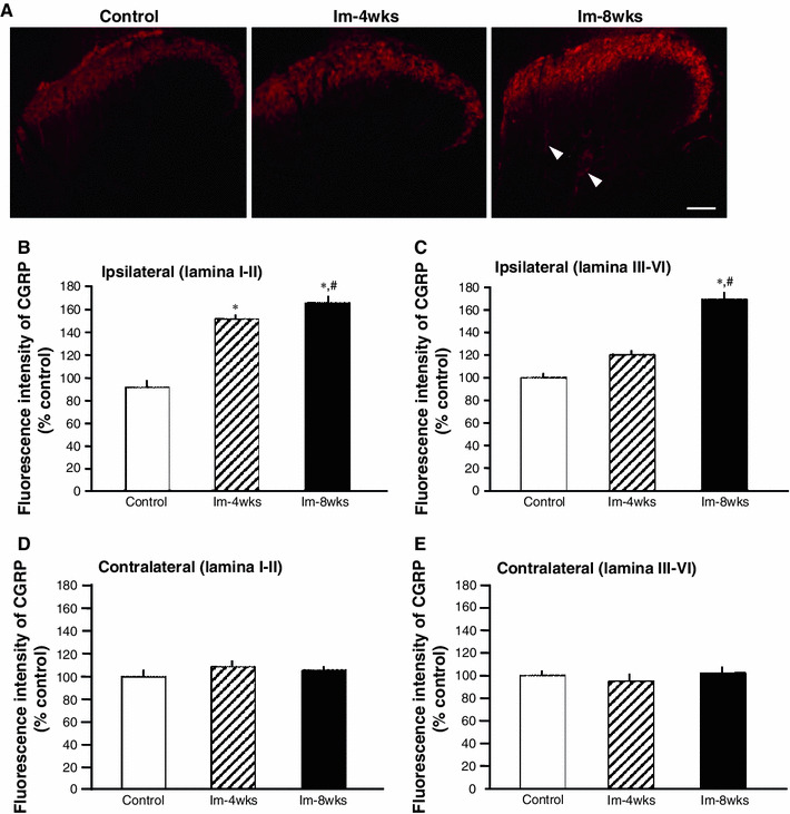
Fig. 4.

Intensity of calcitonin gene-related peptide (CGRP) expression in the ipsilateral dorsal horn of the spinal cord. Representative photomicrographs of CGRP immunohistochemistry in the ipsilateral dorsal horn from the Im-4 weeks, Im-8 weeks, and the control groups (age-matched with the Im-4 weeks group) are shown (a). The CGRP-positive neural fibers were clearly observed in the deep layer of the dorsal horn only in the Im-8 weeks group (arrowheads). Percentage control of fluorescence intensity of CGRP expression in the superficial layer (laminae I-II) (b, d) and deep layers (laminae III-VI) were calculated (c, e) in the ipsilateral and contralateral dorsal horn. *P < 0.05, significantly different compared to the age-matched control group. # P < 0.05, significantly different compared to the Im-4 weeks group. Scale bar = 100 μm
