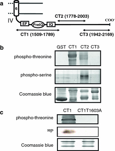
Fig. 1.

Thr1603 of Cav1.2 channel is phosphorylated by CaMKII. a Schematic diagram of the C-terminal tail of the guinea-pig cardiac Cav1.2 channel. EF-hand, preIQ and IQ-motif regions are shown. Fragment peptides, CT1, CT2 and CT3, are shown with a.a. numbers. b CaMKII-mediated phosphorylation of GST-fusion CT1, CT2 and CT3 and GST (negative control) detected with immunoblot blot analysis using anti-phospho-threonine (upper panel) and anti-phospho-serine (middle panel) antibodies. The bottom panel shows Coomassie staining of the gel. c Phosphorylation of CT1 and its mutant CT1T1603A detected by anti-phospho-threonine antibody (upper panel) and autoradiography (middle panel). Coomassie staining is in bottom panel. Each experiment was performed at least three times with equivalent results
