Fig. 7.

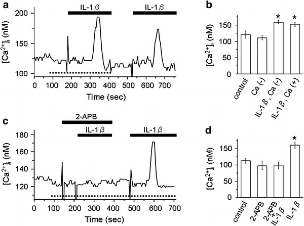
Involvement of intracellular Ca2+ store in the IL-1β-induced rise in [Ca2+]i. a Representative trace of Fura-2 Ca2+ imaging which shows that IL-1β increased [Ca2+]i in both the Ca2+-free bath solution (dotted line at the bottom of trace) and the control bath solution containing 1 mM Ca2+. b Summarized data showing [Ca2+]i under the control condition (n = 10) and [Ca2+]i in response to removal of extracellular Ca2+ (n = 10) and IL-1β with (n = 10) or without (n = 10) extracellular Ca2+. *P < 0.05, significantly different compared with the control. c Representative trace of Fura-2 Ca2+ imaging which shows that 2-APB blocked the IL-1β-induced rise in [Ca2+]i in the absence of extracellular Ca2+. Dotted line at the bottom of trace represents the period during which the Ca2+-free bath solution was used. The doses of 2-APB and IL-1β were 1 μM and 15 pg/ml, respectively. d Summarized data showing [Ca2+]i under the control condition (n = 8) and [Ca2+]i in response to 2-APB (n = 8), IL-1β in the presence of 2-APB (n = 8), and IL-1β alone (n = 8). *P < 0.05, significantly different compared with the control
