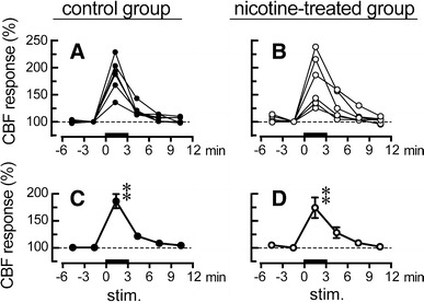
Fig. 5.

Responses of cerebral blood flow (CBF) in the parietal cortex to focal electrical stimulation (at 200 μA) of the unilateral NBM ipsilateral to the parietal cortex in aged rats of the control group and nicotine-treated group. Reponses of CBF during a 3-min period were plotted every 3 min as a percentage of the pre-stimulus values in the saline-treated control group (a, c, closed circles, n = 6) and the nicotine-treated group (b, d, open circles, n = 6). a, b CBF response measured in 6 rats from each group, c, d Summarized graph. Each point and vertical bar represents the mean ± SEM. **P < 0.01; significantly different from pre-stimulus control values (−3 to 0 min) using one-way repeated-measures ANOVA followed by Dunnett’s multiple comparison test
