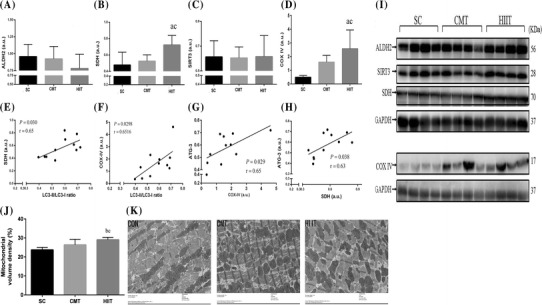
Fig. 3.

Effect of the CMT and HIIT protocols on protein levels of ALDH2 (a), SDH (b), SIRT3 (c), and COX IV (d) in cardiac muscle in the three experimental groups assayed by western blotting (i). Levels of detected proteins were normalized to glyceraldehyde-3-phosphate dehydrogenase (GAPDH). Densitometry analysis was performed to quantify the expression levels of detected proteins (a.u.). Positive correlations of SDH content with LC3-II/LC3-I ratio (e) and ATG-3 (h) in cardiac muscle. Positive correlations of COX IV content with ATG-3 (g) and LC3-II/LC3-I ratio (f) in cardiac muscle. Mitochondrial volume density (j, k) determined using a transmission electron micrograph (at ×6800) of an ultra-thin section of the left ventricle of rat hearts (n = 5). Groups: SC sedentary control, CMT continuous moderate-intensity training, HIIT high-intensity interval training. Data are presented as mean ± SD (n = 4). aP < 0.05 versus SC; bP < 0.01 versus SC; cP < 0.05 versus CMT
